


Core Benefit
This technology enables artifact-free image reconstruction in interior CT, a technique that achieves high-precision CT imaging with significantly reduced radiation exposure. It resolves the fundamental artifact issues that have long hindered interior CT, allowing for clear and accurate images without distortion.
Technology Overview & Background
Unlike conventional CT, interior CT focuses X-ray beams on a limited internal region of the subject, rather than covering the entire body (see right figure). This approach allows for targeted imaging—for example, focusing only on an organ with a tumor or on ischemic regions of the brain or heart—while minimizing radiation exposure to other areas. In industrial CT applications, this technique enables inspection of specific areas (such as defects and foreign matter in semiconductor components) without the need to trim the sample.
Interior CT is a technology that offers significant advantages. However, in interior CT, the method for reconstructing images (calculating cross-sectional images from projection data obtained by X-ray beam irradiation) has not been established, resulting in artifacts (errors) in the CT images obtained. In fact, this issue is not limited to interior CT; it also arises in conventional CT when examining obese patients or large components, as the X-ray beam may not fully cover the target area, leading to a situation similar to that in interior CT. Therefore, this artifact problem can be considered a general challenge that needs to be addressed in CT technology.
To address this, Professor Hiroyuki Kudo and his team at the University of Tsukuba have developed a mathematically rigorous reconstruction algorithm that can significantly reduce or eliminate artifacts in interior CT images. The key innovation is that it requires only a small amount of additional information beyond standard interior CT scan data—without needing to change existing equipment or procedures significantly.
As a result, this technology allows users to fully realize the benefits of interior CT: producing high-resolution, artifact-free images while dramatically lowering radiation exposure.
Data and Publication
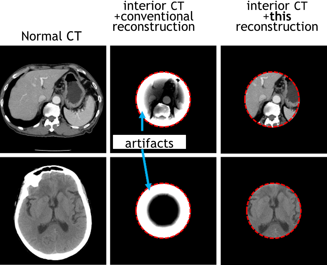
- The research team conducted interior CT scans on human torso and brain samples. When reconstructed using conventional methods, the resulting images were largely unreadable due to heavy artifacts (see center figure). In contrast, the proposed method produced high-accuracy interior CT images that are nearly indistinguishable from those of full-scan CT systems (see right figure). Moreover, the radiation dose required was only 1/4 to 1/8 that of standard CT scans.
 |
Researchers & Academic Institution
Hiroyuki Kudo, PhD (Professor, Faculty of Engineering, Tsukuba University, Japan), et al.
Expectations
We are seeking companies interested in developing and commercializing CT devices (for medical, dental, or industrial non-destructive testing applications) utilizing this technology. Based on the technology provided by Tsukuba University, how about introducing high-precision, low-radiation interior CT image reconstruction? If you are interested in receiving technical details, please contact us. We will arrange a meeting with the developers for further discussion. Regarding technology introduction, we can discuss contracts (for a fee) for the provision of reference programs or lectures by developers.
In addition to interior CT, we also offer image reconstruction technology for sparse view CT. Furthermore, we have high-performance reconstruction technology utilizing compressed sensing and machine learning. Please also refer to the following pages.
- High-Performance Image Reconstruction Algorithm for Sparse Scan CT
- Deep Learning-Based Compressed Sensing Image Reconstruction Technology
Project ID:DA-02355a