


Advantages
- Preservation of the physiological brain environment: Unlike the open-skull method, this technique minimizes inflammatory responses, stabilizes intracranial pressure, and prevents cerebrospinal fluid leakage, enabling long-term, high-precision observation of natural brain activity.
- High-resolution and high-sensitivity imaging: Provides image resolution and signal-to-noise ratios comparable to those achieved with the open-skull method.
- Deep tissue visualization: Allows clear imaging of neurons located deep within the brain (up to 400 µm) through the intact skull.
- Rapid and user-friendly procedure: The skull can be rendered transparent in approximately one hour, enabling immediate initiation of live imaging and significantly enhancing experimental efficiency.
Technology Overview & Background
Conventional brain imaging techniques typically require skull thinning or complete removal, as seen in the thin-skull and open-skull methods. However, these approaches are invasive, may trigger inflammatory responses, and often limit the field of view. In contrast, the “SeeThrough” technique offers a novel, minimally invasive, and high-precision alternative that enables brain observation without bone removal.
Principle and Technical Details
A primary challenge in achieving skull transparency is the bone’s high refractive index (>1.56), which conventional methods have struggled to match.
- Organic solvent–based techniques can achieve sufficient refractive index matching but are highly cytotoxic, making them unsuitable for live imaging.
- Water-based reagents are biocompatible but lack a sufficiently high refractive index to match that of bone, thus limiting transparency.
This new reagent addresses the challenges by offering high biocompatibility and a refractive index sufficiently close to that of bone. Through compound screening and formulation optimization, we have developed a solution that is both practical and effective for long-term live imaging.
Principal Investigator & Academic Institution
Professor Kazuki TAINAKA (Niigata University Brain Research Institute)
Professor Takayasu MIKUNI (Niigata University Brain Research Institute)
Patents
PCT application (unpublished)
Publication(s)
Liu, X., Uchigashima, M., Oomoto, I. et al. SeeThrough: a rationally designed skull clearing technique for in vivo brain imaging. Nat Commun 16, 7584 (2025). https://doi.org/10.1038/s41467-025-62836-1
Current stage of research and future plans
- Using the skull clearing reagent in combination with two-photon microscopy, we achieved clear visualization of neuronal structures that were barely detectable through an untreated Image quality—measured in terms of resolution and signal-to-noise ratio—was comparable to that obtained with the conventional open-skull method.
- No activation of inflammatory markers was observed 24 hours post-treatment, and intracranial pressure remained within normal physiological ranges, confirming the minimal invasiveness of the technique.
- Neural cells were clearly visualized at depths of up to approximately 400 µm, allowing for observation of cortical Layer 5. Furthermore, stable observation was maintained for about one month.
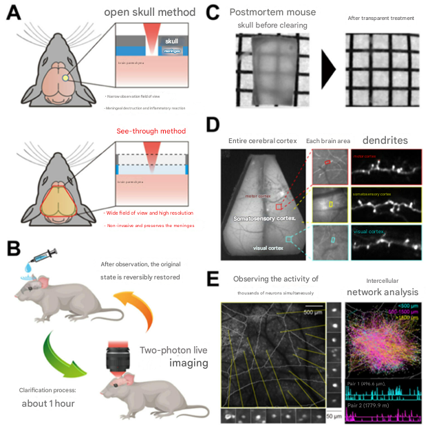 |
Figure Description
(A) Comparison between the SeeThrough method and the conventional open-skull method.
(B) The skull can be rapidly and reversibly cleared for live imaging of the brain.
(C) The mouse skull becomes highly transparent.
(D) Large areas of the skull can be cleared, enabling “zoom-out/zoom-in” observations.
(E) Activity of thousands of neurons across multiple brain regions can be monitored (left), and inter-neuronal network analysis performed (right).
Expectations
TECH MANAGE is actively seeking partners interested in commercializing this technology through kit development and sales, as well as pharmaceutical and microscopy-related companies looking to apply this technology in drug discovery or high-resolution brain imaging.
This technology holds promise for previously unattainable research applications, including:
– Real-time visualization of neural cells under physiological conditions
– Long-term tracking of brain changes in disease model animals
– Observation of immune cells in the glymphatic system and brain border regions
– Drug efficacy evaluation and disease pathology visualization in translational neuroscience
For more detailed information or inquiries, please feel free to contact us. We are also happy to arrange an initial web meeting with the principal investigator upon request.
Project ID:TT-05290