


Advantage and Core Benefit
- Tractography of the lumbar nerve using diffusion tensor images to visualize the site of pain.
- Tractography image creation, which requires experience, time, and cost, to be automated by AI.
- Enables minimally invasive surgery by being able to identify the pain-causing site of lumbar foraminal stenosis, which is difficult to diagnose with imaging.
Background and Technology
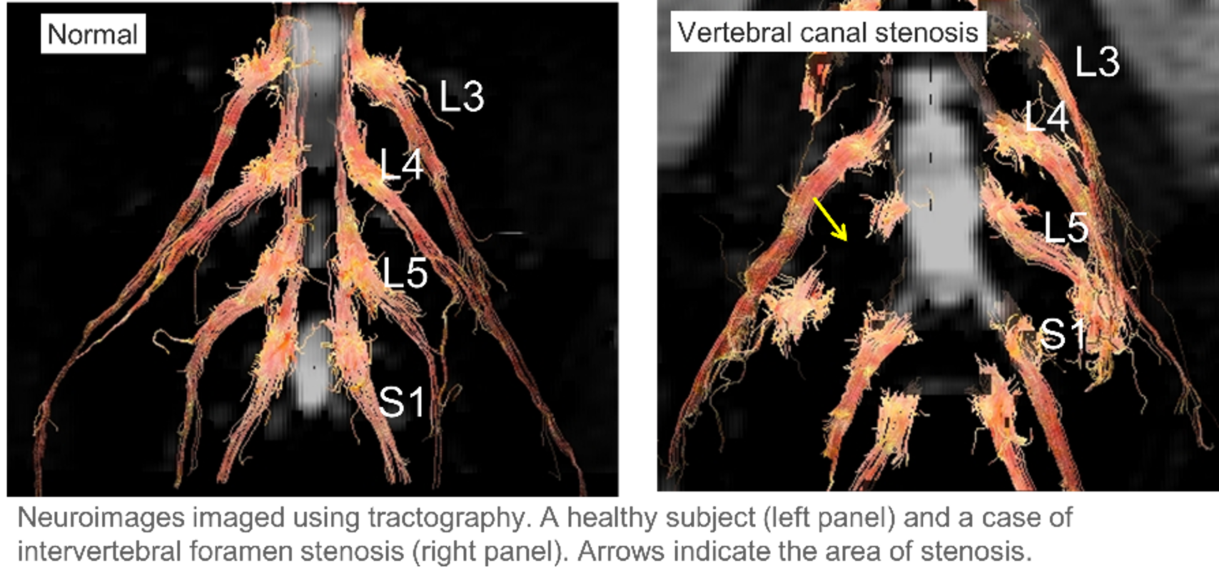
Currently, about 80% of low back pain cases can be diagnosed by detailed MRI imaging. However, there are many false-positive cases in which nerve compression is found on MRI images, but this is not the site of the pain. False positives are extremely high in cases of lumbar foraminal stenosis (40%), making accurate imaging diagnosis difficult. To solve this problem, the inventors reported that the FA value, an index of anisotropy of water molecules in nerve fibers in diffusion tensor images, decreases with nerve damage, and is correlated with clinical symptoms such as pain. We have developed a method to visualize and diagnose pain by imaging lumbar nerve fibers using tractography, a technique to construct nerve fiber runways in three dimensions.
In tractography-based imaging, the stenosis is shown as an image of disrupted nerves. In the imaging process, the nerves had to be manually identified on the FA map, which was problematic in terms of experience, time, and cost. To solve this problem, the inventors developed an automated diagnostic system that identifies nerve roots using AI deep learning segmentation and automatically creates tractography from MRI DICOM data using three free software programs. This allows the system to automatically display and identify the site of the injured nerve using FA values on the tractography, making it possible to visualize and diagnose the site of low back pain in a short period of time without the surgeon’s experience. This will enable minimally invasive surgery that pinpoints the site of low back pain and is expected to reduce patient burden and medical costs.
Data
- The Dice coefficient, an evaluation index, was 0.77 when nerve roots were identified by deep learning segmentation using U-Net.
- We used three free software programs to automatically create tractography from DICOM data, and the average time required was 191 seconds, which was an average of 235 seconds less than the time required
 |
Patent & Publication
- Patent Pending (unpublished)
- Eguchi et.al., Spine Surgery and Related Research 2017; 1(2): 61-71
Researcher
Dr. Yawara Eguchi (Department of Orthopedic Surgery, Chiba University)
Expectations
We are looking for a company that develops image analysis systems that can work with us to develop the automation and universalization of ROI setting and fiber tracking and the visualization of impaired nerves through the licensed introduction of this invention. We can meet with the inventor as well as disclose unpublished data and other information by signing a confidentiality agreement with Chiba University. We can also consider setting up options such as exclusive evaluation for a certain period or preferential negotiation rights for licensing.
Project.WL-04886