


Advantages
- A new technology to measure the charge distribution of a charged particle beam passing at high speed (almost the speed of light) with very high spatial and temporal resolution.
- Expected to improve treatment efficacy and reduce radiation exposure at particle therapy facilities.
- Expected to improve observation and processing accuracy in electron microscopes and focused ion beam systems.
Background
In the realm of accelerators, electron microscopes, and electron beam processing machines such as welding and cutting machines, discerning the three-dimensional shape of charged particle conglomerates is of paramount importance, albeit exceedingly difficult to measure. For instance, in heavy particle therapy, accurately simulating the 3D distribution of charged particle clusters can aid in predicting the behavior of generated particles within a patient’s body, thus improving treatment efficacy, and minimizing undue radiation exposure. Likewise, in electron microscopes, a multitude of electromagnets are utilized to focus the electron beam and correct aberrations, and ascertaining the beam’s three-dimensional distribution is a direct means of validating their intended function.
Regrettably, to date, there exists virtually no non-contact method capable of measuring the distribution of charged particles moving at nearly the speed of light as they pass through the beam. Previously proposed technologies have proven inadequate in terms of providing sufficient spatial resolution.
Technology
Associate Professor Makoto Nakajima of the Institute of Laser Engineering, Osaka University, has developed a new technique for measuring the charge distribution of charged particles with high spatial resolution.
The principle of this technique is to measure the change in birefringence (Pockels effect) that occurs in an electro-optic crystal. When an object to be measured (a lump of charged particles) passes in the vicinity of an electro-optic crystal, the refractive index in the crystal changes due to the electric field of the charged particles. This change in refractive index perturbs the laser light irradiated onto the electro-optic crystal. The perturbation of the laser beam is read out by an optical sensor to measure the time variation of the charged particle-derived electric field, and the charge distribution of the charged particles is reconstructed based on this time variation. This technique has existed for some time, but because it can only measure the charged particles at the instant when they pass in front of the crystal, it only measures one section of the particle mass and cannot measure the entire mass. This technique solves this problem. By applying a trick to the laser beam, the developers have made it possible to intermittently measure a cross-section of a particle clump and to measure the shape of the entire clump. The trick is to make the laser beam reflect off an echelon mirror. The reflected laser light is not one and the same light but contains multiple rays of light with time differences in their bundles. Each of the rays in the bundle enters the electro-optic crystal with a slight time delay. Then, as each ray enters the crystal, it is perturbed by refraction from the particle mass and exits the crystal. Finally, each ray is focused by a cylindrical lens and detected by an optical sensor. Each light beam has cross-sectional information on the cloud of particles at the timing of its incidence on the crystal. In other words, the cross-sectional information of each light beam enables us to determine the overall shape of the cloud of particles.
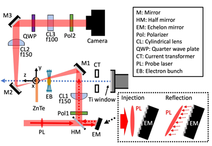 |
Figure 1. Overview of the measurement system for this technology. The electromagnetic field produced by the electron beam (EB, shown in the center of the figure) changes the refractive index of the electro-optic crystal (ZnTe). This change in refractive index perturbs the laser light (shown as the red band) incident on the ZnTe. The measurement of this perturbation with the Camera reveals the distribution of EB. When the incident laser light is reflected by the echelon mirror (EM, shown in the lower part of the figure), it is separated into rays slightly delayed. As a result, the laser beam becomes a group of slightly delayed beams. When this group of light is continuously incident on the electro-optic crystal, the device can measure the distribution of EBs continuously in time. (Nature Phys. volume 18, pages1436–1440 (2022))
Expectation
Osaka University is looking for a company to license this technology and develop a device to measure the charge distribution of charged particles. By signing a partnership agreement with the University, the University can provide the necessary know-how for product development and establish a joint development framework.
We are also seeking information about the needs of electron microscope manufacturers and companies that use charged particle beams, such as focused ion beam instruments. By signing a partnership agreement with a university, you can co-develop equipment to solve your challenges.
Application
For Medical.
Calibration of particle beams used in heavy particle therapy. The precise geometry of the particle beam provided by this technology improves the accuracy of pre-treatment radiation exposure simulation. The simulation results provide a highly accurate reproduction of the dynamics of the particle beam in and around the diseased area. The simulation results provide important information for improving treatment efficacy and reducing radiation exposure.
For Engineering.
・Calibration of particle beams used by electron microscopes, focused ion beam (FIB), electron beam welding and impurity doping equipment in semiconductor manufacturing. This technology improves the accuracy of micro-fabrication.
・Measurement of the charge of the sample in FIB and electron microscopy. The application of this technology is a device for measuring the charge state of objects. This device facilitates the development of countermeasure techniques against defects caused by charge-up. Such defects include the destruction of semiconductor circuits due to short circuits and the blurring and misalignment of microscope images.
Publication
The following article describes the technology and reports on experiments using it. The developers of this technology were the first in the world to measure certain phenomena predicted by special relativity.
https://doi.org/10.1038/s41567-022-01767-w
Project No. DA-03928